

Justin Lim ’26
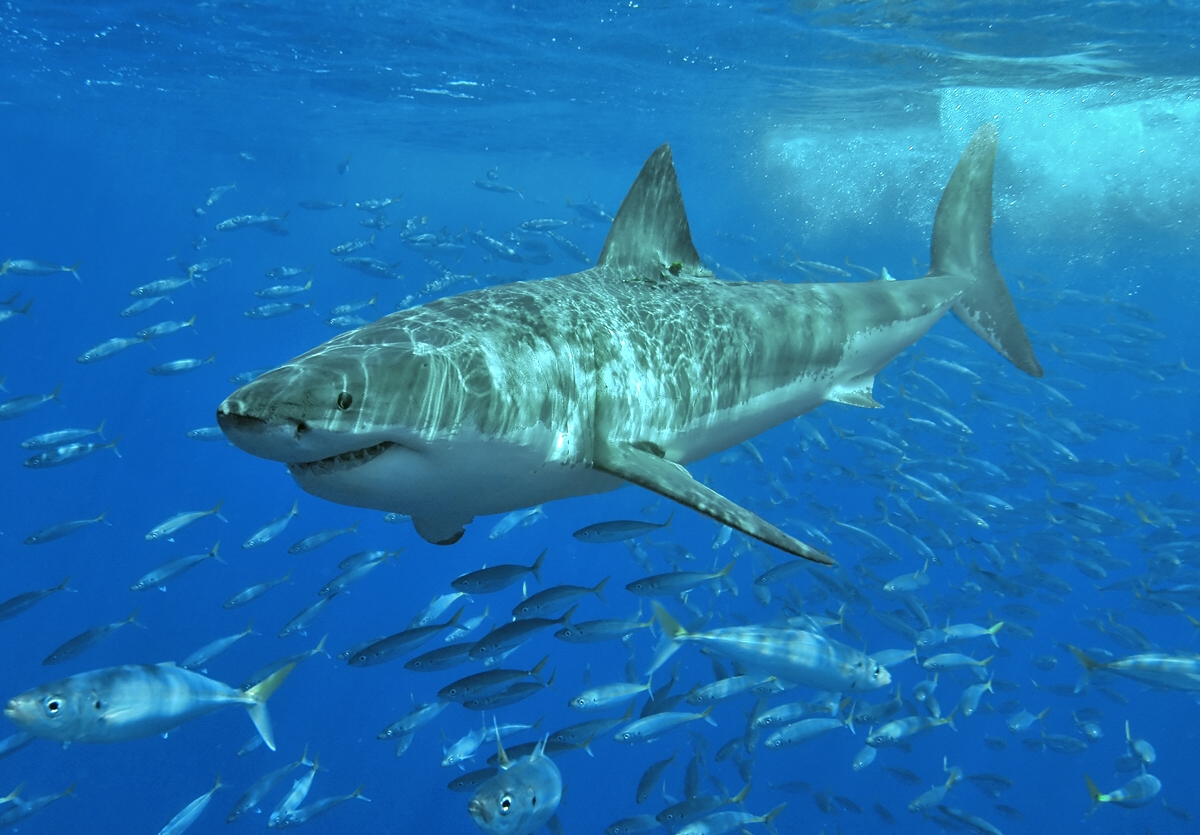
Figure 1: White shark.jpg
Urea, a common byproduct of protein metabolism, is excreted through urine in most urea-producing organisms due to its toxic properties in high concentrations. Specifically, urea’s nitrogen content can destabilize the structure of proteins by weakening their folded structure. Sharks have chemical countermeasures to halt the progression of protein denaturation through the use of trymethylamine N-Oxide (TMAO), a molecule known to stabilize proteins in environments high in urea concentration. However, Satoshi Kanoh and his team at Mie University of Japan hypothesized that shark muscle cells – mostly consisting of protein molecules – have intrinsic resistance to urea. As such, they investigated the urea-resistibility of shark skeletal muscle myosin, the group of motor proteins responsible for muscle contraction, in the absence of TMAO.
Kanoh and his team ran a comparative study between carp and hound shark light myosin chains. They treated shark and carp A1-LC regions of their light myosin chains with 0–2.0 M urea, and then analyzed the spectral profiles of the treated samples at room temperature using a CD spectropolarimeter. The primary purpose of the experiments was to evaluate the alpha-helical concentration of certain regions of shark and carp peptide chains (residues 28–34). Since the concentration of alpha-helices and the structural integrity of proteins are directly related, the already-known protein denaturation effect caused by high concentrations of urea can be utilized to discriminate between the two myosin types shown in the study. The researchers found that as urea concentration increased, carp light myosin chains lost their alpha-helical concentration, therefore displaying protein denaturation. In contrast, the alpha-helical content of houndsharks in the same A1-LC region remained unchanged or increased. The experiment results suggest that shark myosin exhibits urea-resistibility regardless of the urea-TMAO counteraction mechanism.
The study found that shark myosin exhibits native activity at physiological urea concentrations even without TMAO and functions properly even under conditions that usually denature proteins of other animals. These findings provide insight into fundamental aspects of protein stability; insights into how shark myosin maintains structure and function in the presence of urea could have broad applications, ranging from therapeutics to biotechnological advancements. This research not only opens avenues for preventing protein breakdown in human cells, with potential implications for various medical conditions, but also sparks optimism for future breakthroughs in addressing these health challenges.
Source: Kanoh, S., Noma, T., Ito, H. et al. Myosin light chain of shark fast skeletal muscle exhibits intrinsic urea-resistibility. Sci Rep 13, 4909 (2023). https://doi.org/10.1038/s41598-023-32228-w
Image: https://commons.wikimedia.org/wiki/File:White_shark.jpg
