
Author: Amal Bilal, Class of 2028
Chronic wounds, or wounds that demonstrate abnormal repair due to a deficient healing process, pose major healthcare challenges worldwide. These wounds can lead to pain, infection, hospitalization, surgery, and, in the worst cases, amputation. Therefore, early identification of whether a wound is healing properly is crucial for adjusting treatment plans, reducing side effects and costs, and ultimately improving patient outcomes. Tengfei Ma, a biomedical informatics researcher at Stony Brook University, and his team explored the potential of quantitative ultrasound imaging and machine learning to track the wound healing process. By analyzing ultrasound images at different stages of healing, the team aimed to create a machine learning model that can predict wound status and detect abnormalities in healing earlier than traditional methods.
To accomplish this task, the researchers created small wounds on laboratory mice, recorded observations and measurements of their wound healing process, and imaged them over three weeks using standard B-mode ultrasound equipment. Specifically, the diameter and depth of the wound, and features such as pixel brightness and texture within the wound bed and surrounding tissue. These ultrasound findings were subsequently compared to histological tissue data, which showed changes in collagen and cell growth during the healing process. Lastly, the team trained machine learning models, including a pretrained deep learning model called ResNet, which was used on the ultrasound images and extracted features to classify the different stages of healing.
The analysis showed that ultrasounds could reliably track wound closure, since measurements closely matched the actual wound size as healing occurred. For instance, certain image features like brightness peaks and tissue echogenicity (how well tissue reflects ultrasound waves) correlated strongly with biological changes in healing. Moreover, the machine learning models were able to classify the three major healing phases of inflammation, proliferation, and maturation with the best-performing ResNet-18 model achieving 89.7% accuracy and an F1-score of 0.87 in classifying inflammation, proliferation, and remodeling phases. Essentially, the study found that automated methods for image analysis performed similarly to manual analysis, reducing the time and energy required for clinicians to perform the same task.
Overall, the research shows that the combination of ultrasound technology and machine learning offers a powerful, non-invasive, and accessible way to monitor wound healing. This method enables earlier detection of problems with chronic wounds, leading to more personalized treatment and improved patient outcomes. Future work will aim to include a wider range of wound types, enhance ultrasound technologies for continuous monitoring, and apply these findings from mice in human clinical settings.
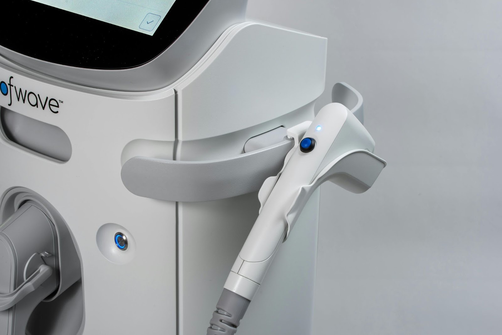
Figure 1, Ultrasound machine.
Works Cited:
[1] Ma, T., Lemonnier, D., Sumpio,. B., et al. “Quantitative Ultrasound Augmented with Machine Learning to Assess Tissue Microstructure during Wound Healing.” Biomedical Signal Processing and Control, vol. 103, May 2025. https://doi.org/10.1016/j.bspc.2024.107420.
[2] Image retrieved from: https://www.pexels.com/photo/a-selective-shot-of-an-ultrasound-machine-9973861/
