

Gaurav Sharma ’22
An important part of development of any organism is differential regulation of the cell cycle since it leads to cell specification and differentiation. The cell cycle states and their coordination are already well-studied, but the mechanistic connection between the cell cycle and differentiated cell behavior is still to be determined. The challenge is finding a reliable live cell imaging tool that can accurately characterize cell cycle state and preserve the normal processes in the cell. One indicator used is fluorescent, ubiquitination-based cell cycle indicator (FUCCI) which relies on degrading specific DNA factors. The drawbacks of it are that it fails to image cells with fast cell cycles and/or translational activity as well as not clearly distinguishing the S and G2 phases. The Matus Lab at Stony Brook University sought out a novel method to visualize cell cycle states in C. Elegans to solve these issues.
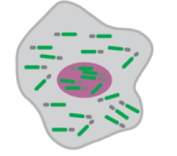
Researchers in the Matus Lab decided to look for a phosphorylation biosensor rather than a degradation biosensor. Building off of previous biosensors using cyclin-dependent kinases (CDKs), the Matus Lab created an optimized version of DNA Helicase B (DHB) fused to a green fluorescent protein (GFP) and a histone. Using the CRISPR/Cas9-genome engineering, a CDK sensor was inserted. Together, fluorescence in the nucleus and cytosol could be observed and compared to precisely determine the cell cycle state that the cell is currently in. The range of the biosensor was also tested and it showed reliability under low nuclear expression. Different strains were made to balance fluorescence, photobleaching, and mitotic exit.
These findings offer a novel method for observing cell cycle state in C. Elegans which can be used for other Metazoa. This method is important for observing how differentiation occurs in conjunction with the cell cycle. It could also be used to find other connections to the cell cycle such as the role/control of the cell cycle and tissue regeneration or cell cycle defects and developmental defects. Currently, the Matus Lab is looking more into cell cycle activity and basement membrane invasion.
[1] A. Kohrman et al. Visualizing the metazoan proliferation-differentiation decision in vivo.
BioRxIV. 1-37 (2019). doi: https://doi.org/10.1101/2019.12.18.881888
[2] Image retrieved from: https://www.biorxiv.org/content/10.1101/2019.12.18.881888v1.full.pdf
