
By Adil Ali Khan, Class of 2029
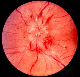
Figure 1: Papilledema, a form of optic disc edema (swollen optic nerve), often mimicking bilateral optic disk swelling, which is characteristic of Vogt-Koyanagi-Harada disease
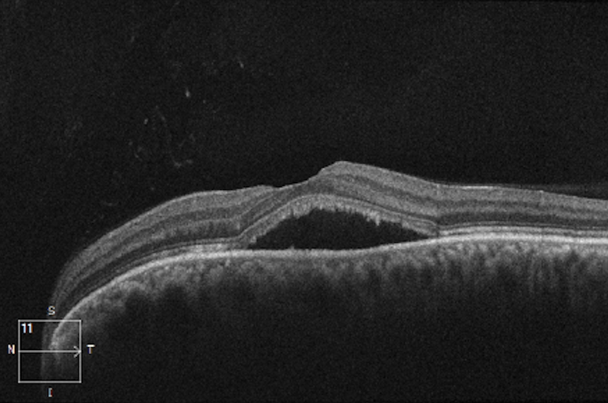
Figure 2: Macular OCT depicting serous retinal detachment in the left eye, a symptom of Vogt-Koyanagi-Harada disease
Vogt-Koyanagi-Harada (VKH) disease is a systemic inflammatory condition characterized by autoimmune responses that target melanocytes and affect multiple organs, including the eyes, skin, inner ear, and meninges. Ocular symptoms include bilateral panuveitis (inflammation throughout the uvea in both eyes), serous retinal detachment (SRD; retina peels away from the back of the eyes), and optic disc edema (swollen optic nerve). Current treatment for VKH disease leads to favorable visual acuity outcomes, but the exact recovery process of visual functions – color vision specifically – remains incompletely understood.
Cone cells (or cones) are responsible for color perception in human eyes. This research was conducted by the Department of Ophthalmology at Toyama University Hospital in Japan. The researchers aimed to use cone contrast testing to evaluate qualitative aspects of visual function that conventional visual acuity tests do not capture. This was done by individually assessing L-cone (long-wavelength-sensitive), M-cone (medium-wavelength-sensitive), and S-cone (short-wavelength-sensitive) functions and by detecting color vision abnormalities in various ocular diseases. The researchers also used adaptive optics (AO) technology to obtain high-resolution images of cone cells in human eyes. It was hypothesized that VKH disease initially reduces cone cell density, but treatment allows the density to increase over time.
The study found that at the time of SRD resolution, S-cones demonstrated clear color-vision abnormalities, whereas L- and M-cones exhibited potential color-vision deficiencies. After 3 months of treatment, however, L- and M-cone functions returned to normal ranges. After 6 months, notable progress was observed across all cone types, reaching standard ranges, indicating that the impact of VKH disease on color vision is temporary, with substantial recovery feasible within approximately 6 months. The research also found that the S-cones were more vulnerable and recovered more slowly than L- and M-cones. Evidence from glaucoma and other inflammatory diseases supports the finding that S-cones are more susceptible to damage. Using AO imaging, the researchers were able to visualize clear structural changes in the cone photoreceptor layer that correlate with functional color vision deficiencies. They were particularly able to observe the reduction in cone cell density, demonstrating a strong structure-function relationship. Treatment resulted in recovery of cone density, again supporting the fact that VKH disease causes reversible structural damage to the cone cells.
This study is especially important because it examines how color vision is recovered in VKH disease, an under-researched condition. Further investigating the factors that influence S-cone recovery would provide a deeper understanding of color vision restoration and potentially inform treatments for other retinal conditions.
Work’s Cited:
[1] Nakamura, T., Abe, S., Yamazaki, H., Oiwake, T., & Hayashi, A. (2025). Dynamics of color vision recovery in Vogt-Koyanagi-Harada disease: a longitudinal study using cone contrast test and adaptive optics imaging. Journal of ophthalmic inflammation and infection, 15(1), 64. https://doi.org/10.1186/s12348-025-00523-4
[2] Image (Figure 1) retrieved from: https://commons.wikimedia.org/wiki/File:Papilledema.jpg
[3] Image (Figure 2) retrieved from: https://commons.wikimedia.org/wiki/File:Macular_OCT_depicting_Central_Serous_Chorioretinopathy_in_the_Left_Eye.png


