

By Adil Ali Khan, Class of 2029
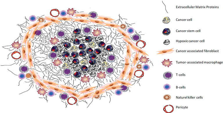
Figure 1: Schematic view of the tumor microenvironment
Polyploid giant cancer cells (PGGCs), found in tumors, have long been dismissed as excessive inflammatory waste with no biological or clinical relevance. Yet recently, numerous researchers have described the existence of PGGCs in solid tumor masses that appear to correlate with tumor progression, also appearing as cancer-associated macrophage-like cells (CAMLs) in the blood circulation. To understand the biological and clinical function of CAMLs, which are PGGCs in circulation, researchers at SBU initiated a multi-institutional 2-year prospective study of patients with a range of solid tumors.
Scientists at several major US cancer research centers used CellSieve size-based low-flow microfiltration to capture cells and isolate CAMLs. These CAMLs were identified based on size, polyploid nuclei, and expressions of four stains: nuclear, epithelial, leukocyte, and myeloid markers. Next, the polyploidy status of the CAMLs was verified using a fluorescence-based molecular tool, the CEP17 FISH probe, which detects polyploidy by the number of chromosome 17 copies in a cell’s nucleus. Sequential QUAS-R (Quench, Underivatize, Amine-Strip, and Restain), a chemical imaging technique, was used to remove fluorescence from signals from the tumor and cells to allow for re-staining with new antibodies and identifying which markers were visualized. The cells were cultured in vitro to evaluate proliferation and behavior. Lastly, statistical analyses, such as Kaplan-Meier curves and hazard ratios, assessed the relationships between CAML size and progression-free survival (PFS) and overall survival (OS).
The study’s findings showed that CAMLs were detected in 93% of cancer patients, with higher proportions in more severe cancer stages than in less severe ones, and were absent in all healthy controls. Moreover, it showed that larger CAMLs, those with higher ploidy, were associated with substantially poorer PFS and OS, irrespective of cancer stage. In terms of phenotyping, CAMLs co-expressed markers across lineages, including myeloid, epithelial, endothelial, and stem, as well as motility receptors. Many displayed combinations not typical of normal differentiation, suggesting complex origins such as phagocytosis of tumor material. However, a proportion of CAMLs adhered to typical proliferation in culture, consistent with stem-like activity.
Overall, the study clearly showed that CAMLs were prevalent even in early stages of cancer, unlike typical circulating tumor cells, and were associated with progression before metastasis. This study is the most detailed examination of circulating PGCCs to date, and yields results that refute misconceptions about CAMLs, such as their biological irrelevance. These findings highlight CAMLs as promising, non-invasive biomarkers that can help track cancer progression and better predict patient outcomes.
Work’s Cited:
[1] Adams, D. L., Cristofanilli, M., Lin, S. H., Bergan, R. C., Ho, T. H., Marks, J. R., Martin, S. S., Edelman, M. J., Chumsri, S., Hager, E. J., Tang, C. M., Tsai, S., & Alpaugh, R. K. (2025). Phenotyping and clinical utility of phagocytic polyploid giant cancer macrophages in blood. Cancer Letters, 631, 218007. https://doi.org/10.1016/j.canlet.2025.218007
[2] Image retrieved from: https://commons.wikimedia.org/wiki/File:A_schematic_view_of_the_tumor_microenvironment.png


