

By Sajia Athai Class of 2026
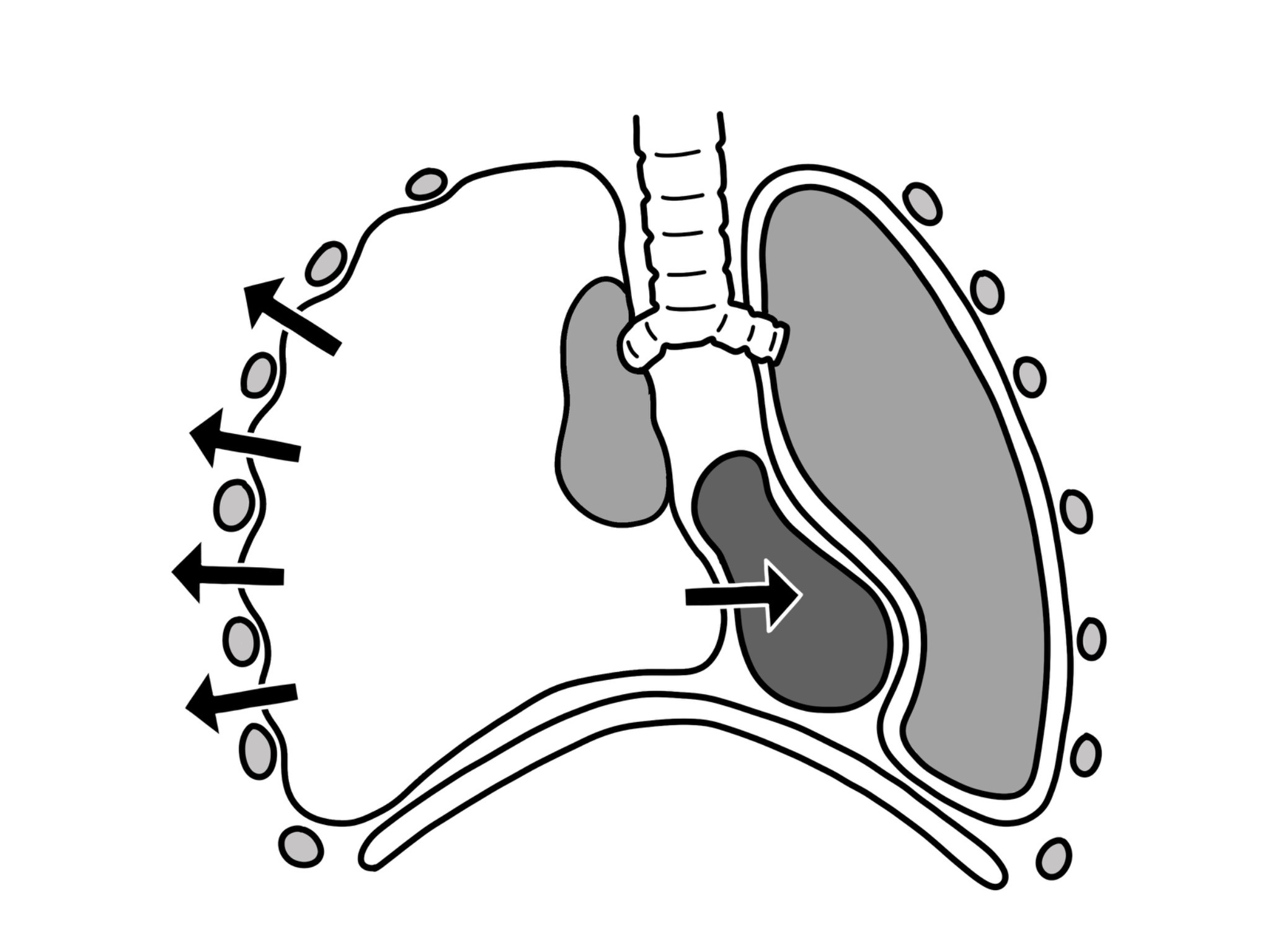
Figure 1 Pneumothorax (PTX), the collapse of a lung, is an integral condition that existing techniques in the past have incorrectly diagnosed. New advances show more accurate diagnosis methods, highlighting patient sensitivity and responsiveness.
In trauma patients, there are established techniques that are utilized to detect pneumothorax (a collapsed lung), typically consisting of a supine chest x-ray. In a supine x-ray, the patient is lying down while an x-ray is performed, making certain organs, such as muscle, look larger in size. Supine x-rays can make it difficult to detect small pneumothorax (PTX) , making it more challenging for doctors to treat and diagnose trauma patients. Recently, a group of surgeons and researchers at the Stony Brook Renaissance School of Medicine, led by Dr. Isadora Botinick, conducted a study on a technique that would allow them to more effectively use thoracic point-of-care ultrasound, also known as POCUS, as a diagnostic technique for PTX. While this new discovery exhibits significant potential in effectively reducing errors in detecting PTX, new research raises questions about its clinical relevance in the diagnosis of patients.
The researchers observed a sample size of 924 patients over three years in a level I trauma center. Through tomography scans and clinical descriptions made based on tube thoracostomy placement, the diagnosis of PTX was either made or ruled out. By measuring the amount of time a patient required a tube thoracostomy, the scientists established clinically significant standards pertaining to this time of triage of within 2.5 hours. The supine x-ray and the point-of-care sound studies were then conducted to compare statistical conclusions. Out of the 924 patients, 40 had clinically significant PTX, revealed by the point-of-care ultrasound method. 26 out of 38 patients—who survived before tomography scans were conducted—exhibited a 68% sensitivity rate. This point-of-care scan technique yielded a sensitivity improvement of about 84% while the overall sensitivity in the use of supine x-ray yielded an overall percentage of around 48%. The tremendous difference in sensitivity indicates the effectiveness of using point-of-care ultrasounds in diagnosing and detecting PTX in comparison to established methods, especially for clinically significant PTX. While the images produced will need a more refined skillset in interpretation and identification, the technique promotes greater accuracy in the detection of PTX in patients.
With this major advancement in surgery and emergency medicine, medical care professionals hope the technique can be implemented in hospitals globally to minimize the risk of incorrectly diagnosing a patient and neglecting PTX treatment.
Work’s Cited:
[1] Singer D. D., Scott H., Khan A., Donnelly A., Singer A. J., Botwinick I., Jawa R., Mukhi A., Thode H. C., & Secko M. (2025).Emergency Department Accuracy of Point-of-Care Ultrasound in Identifying Clinically Significant Pneumothorax in High-Severity Trauma Patients. The Journal of Emergency Medicine, 77,, 140-151. https://doi.org/10.1016/j.jemermed.2025.07.009
[2] Image retrieved from https://commons.wikimedia.org/wiki/File:Tension-pneumothorax.png


