

By Sajia Athai, Class of 2026
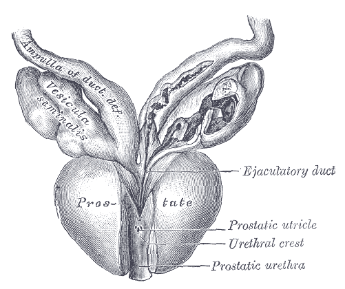
Figure 1 The U-Found software tool was designed to detect cancer in the prostate based on analysis of the entire gland, rather than a few regions of interest.
Dr. Joel Saltz, the Founding Chair of the Department of Biomedical Informatics at Stony Brook University, is part of a team of researchers who have developed a new AI software model to analyze prostate MRI scans. While various current tools solely focus on the detection of tumors in the prostate, the U-Found analyzes the entire prostate gland and its surrounding environment. The research team strongly believes that to advance cancer imaging detection methods, techniques used to track progression of cancer should be expanded beyond a targeted region of the gland to maximize chances of recovery.
Men who are diagnosed with low-risk prostate cancer often choose Active Surveillance (AS), a set of preventative and diagnostic tests aimed at monitoring the progression without immediate treatment. However, due to patients experiencing different symptoms and progression markers, AS is not an ideal medical option for all. In addition, numerous biopsies were often required to monitor progress—resulting in patient discomfort and delays in progression analysis. U-Found, on the other hand, is a foundational tool that represents MRI images as 128-dimensional vectors that identifies patterns across different prostate scans, essentially outlining regions of interest among prostates. Utilizing data from 3,244 patients, approximately 49,000 MRI slices were used to train the model to recognize patterns without any information about tumor location in the prostate. The goal is for the software to be able to distinguish between similar and disparate images, which was implemented through contrastive learning (SimCLR). By combining the embeddings from MRI scans with clinical patient data, the software model can predict progression markers in AS patients diagnosed with prostate cancer. The images captured by the software display indications of features that are ideal for observation in the prostate.
Utilizing embeddings to predict cancer, the software integrates logistic regression measures, labeling the image slices made from the data as “cancer” or “no cancer”. Measuring the accuracy of the detection markers indicated by the software, the AUC (Area Under the Curve) measure was found to be 0.79, affirming the reliability of the tool. This technique can be utilized to mark the progression of cancer more quickly and efficiently in patients without causing discomfort and delays in treatment.
Work’s Cited:
[1] N. C. Lowry, et al.,MRI-based patient selection for active surveillance in prostate cancer using U-Found: A generalized deep learning model. Cancer Imaging 26, 1 (2026). doi: 10.1186/s40644-026-00988-z.
[2] Image retrieved from https://garystockbridge617.getarchive.net/amp/media/prostate-gray1153-4b8d9d.
https://commons.wikimedia.org/wiki/File:Prostate_-_Gray1153.png


