

Aaradhana Natarajan, 2020
The Human Immunodeficiency Virus, commonly called HIV, is one of the most widespread immune disorders across the globe. It weakens the immune system by reducing lymphocyte count and increasing susceptibility to other diseases. While it is most commonly known for its transmissibility, it is also possible for HIV-positive pregnant women to pass on the infection to their offspring. This form of HIV is known as perinatally acquired HIV (PHIV). While its immunological impact is well known, researchers at Northwestern University were interested in studying the virus’s neurological effects. Led by Dr. Paula Lewis-de los Angeles, the team investigated the connections between PHIV severity and grey matter volumes in the brain.
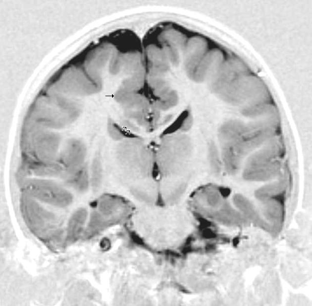
The researchers gathered a PHIV-positive cohort from the NIH Pediatric HIV/Aids Cohort Study network, and a control group from the Pediatric Imaging, Neurocognition and Genetics study. Both groups were frequency-matched for sex and age. To determine PHIV severity, each individual was tested for CD$ T-lymphocye oercentages (CD4%) and plasma HIV RNA concentrations. The former was used to determine disease severity, and the latter to determine viral load. They also recorded the lowest known lifetime CD4% and highest known lifetime RNA concentrationCortical and subcortical grey matter volumes were measured for certain regions of interest (ROIs) using structural MRIs.
The results indicated that PHIV infected youth had significantly smaller volumes in the ten primary ROIs. Peak VL load also negatively correlated with total grey matter, suggesting that increased PHIV severity corresponded to lower volumes. Higher VL also correlated with reduced grey matter in the bilateral rostral middle and superior frontal lobes. These results were sustained even after adjustment for sex, age, caregiver educational attainment, race, annual household income and intracranial volume.
On a side note, the researchers also measured substance abuse was measured through electronic self-interviews. The results indicated that PHIV-positive individuals who reported alcohol or marijuana use also had total volumes that were anywhere from 9-16% smaller than PHIV-positive individuals who reported no drug use.
In analyzing these results, it is important to consider that there has not been extensive research into the effects of long-term HIV infection on adolescent brain development. To this end, it is not certain whether it is the drug consumption that fosters reduced volume and aberrant neurological development, or the other way around. The researchers did not compare the data for VL, RNA levels and drug consumption, which may prove a future area of interest. Similarly, the correlation between inflammatory markers and PHIV severity is still relatively unknown.
Limitations include the use of only a single neuroimaging visual, taken during the study. This prevented the researchers from understanding the longitudinal impact of PHIV on neuroanatomical development. Also, the examination of drug use was based on whether or not the individuals had ever used, rather than based on frequency or duration of use. Despite these difficulties, however, research into the neurological effects of immunological disorders, particularly one as pervasive as HIV, can foster improved clinical understanding of long-term effects. It can also lead to improved therapies to mitigate the adverse effects of PHIV on growing minds.
Sources:
- Lewis-de los Angeles, C. P., et al. (2017). Lower total and regional grey matter brain volumes in youth with perinatally-acquired HIV infection: Associations with HIV disease severity, substance use, and cognition. Brain, behavior, and immunity, 62, 100-109.
- 2. Image retrieved from: https://commons.wikimedia.org/wiki/File:Grey_matter_heterotopia_MRI.jpg
