

By Caleb Sooknanan ’20

Viral central nervous system (CNS) infections are often associated with cerebrospinal fluid (CSF) lymphocytic pleocytosis, or an increase in the fluid’s white blood cell count. However, these infections are also connected to neutrophilic pleocytosis, an increase in the fluid’s neutrophil count. The clinical and prognostic significance of CSF neutrophilic pleocytosis remains unknown in patients. Doctor Siraya Jaijakul and researchers at the University of Texas Health Science Center at Houston conducted a study to compare the clinical and laboratory characteristics of viral CNS infections with a CSF neutrophilic pleocytosis to those with a lymphocytic pleocytosis. The study also enabled the researchers to understand the adverse clinical outcomes connected to neutrophilic pleocytosis.
The study sample comprised 182 patients with viral CNS infections — as confirmed via CSF culture, serology, or polymerase chain reaction (PCR) — who were evaluated in Houston, Texas and New Orleans, Louisiana from 1999 to 2013. The researchers defined CSF neutrophilic pleocytosis as a CSF neutrophil count more than 50%, while CSF lymphocytic pleocytosis was defined as a CSF lymphocyte and monocyte count of over 50%. The etiologies of the patients’ cases were divided into enterovirus, herpes virus, and arbovirus cases based on whether CSF PCR or viral culture was positive, as well as whether serum or CSF Immunoglobulin G (IgG) or Immunoglobulin M (IgM) was positive for the West Nile Virus (WNV) or Saint Louis Encephalitis Virus (SLEV).
Of the 182 chosen patients, 74 patients had an enterovirus infection, 72 had a herpes virus infection, and 36 had an arbovirus infection. 25% of the patients were designated as having a CSF neutrophilic predominance, while the other 75% had a CSF lymphocytic predominance. Enterovirus infections elicited 64% of neutrophil-predominant CSF and 33% of lymphocyte-predominant CSF, while herpes infections were the cause of 46% of lymphocytic pleocytosis and 20% of neutrophilic pleocytosis. With this, the researchers found that neutrophilic pleocytosis was more common in younger patients, patients with respiratory symptoms, and patients with higher CSF white cell counts.
Limitations of this study included the small sample size of the neutrophilic pleocytosis group, which may have limited the data on repeated CSF study results and the observations of pleocytosis in patients. Additionally, there were differences in sensitivity and specificity between the diagnostic methods used over the study period, so the results may have been affected by diagnostic bias among the available patients. Nevertheless, the study showed that CSF neutrophilic pleocytosis were connected to viral CNS infections. More research is needed with larger sample sizes to support these results.
References:
- S. Jaijakul, et al., The clinical significance of neutrophilic pleocytosis in cerebrospinal fluid in patients with viral central nervous system infections. International Journal of Infectious Diseases 59, 77-81 (2017). doi: 10.1016/j.ijid.2017.04.010.
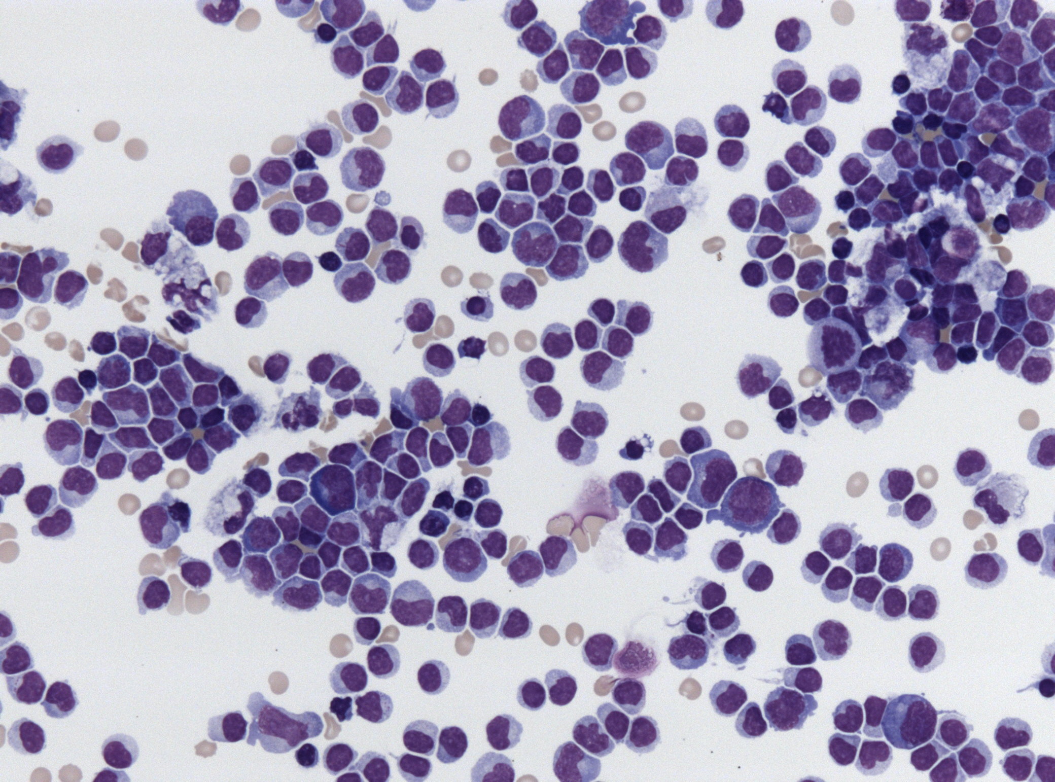
- Image retrieved from: https://upload.wikimedia.org/wikipedia/commons/a/af/Hiv_meningeoencephalitis_csf_pleocytosis.jpg
