

Vignesh Subramanian ’24
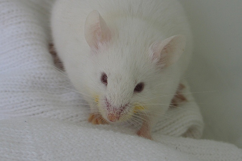
In the central nervous system (CNS) and peripheral nervous system (PNS), glia are supportive cells that form myelin sheaths, or coatings, that insulate and protect neurons. Activated glial cells are also capable of producing growth factors such as BDNF and bFGF that trigger neuroinflammation, inducing a prolonged state of pain which alerts an organism to potential nerve injury. As such, various subtypes of glial cells are clinically viable for therapies targeting neurodegenerative diseases that involve demyelination, such as multiple sclerosis (MS). However, transplantation of these glial progenitor cells – capable of differentiating into specialized subtypes – is often rejected by the mammalian immune system, which recognizes the cells as foreign. To overcome this challenge, a study conducted at Johns Hopkins University explored an approach by which glial-restricted progenitor (GRP) allografts could be transplanted into mice without the use of anti-rejection drugs that a human patient would have to remain on indefinitely.
Researchers used two antibodies – CTLA4-Ig and anti-CD154 – to manipulate the costimulatory blockade-based signals that typically activate an immune response. These antibodies were injected intra- and post-operatively into the brains of three groups of demyelinated shiverer mice to block activation of immune T cells. Researchers also tested a set of five plasma microRNAs (miRNAs), small molecules that can regulate the expression of secreted T cell proteins, for their ability to surveil the graft’s histocompatibility. Bioluminescent and magnetic resonance imaging found that the glial progenitor cells’ fluorescent tags lasted long after transplantation and that there were no significant structural differences between the mice brains, suggesting that the GRP cells survived abundantly enough at specific localizations to produce the necessary sheaths. RNA isolation procedures revealed that the miRNAs were upregulated in control mice when grafts were rejected and downregulated in mice treated with antibodies, suggesting they were capable of accurately tracking the T cell activation cascade that triggered immune responses.
This study demonstrated a successful prevention of graft rejection while obviating the negative effects of immunosuppression protocols, and was the first of its kind to use this antibody combination to repair myelin in the brain, which had not previously been accomplished. The study further established that miRNAs could serve as biomarkers of transplanted cells due to their greater tissue specificity, addressing a long-standing barrier for translation of cell-based therapies. Such developments show great promise for future immunomodulatory treatments designed to protect myelin formation and greater neuronal integrity.
Works Cited:
[1] S. Li, et al., Induction of immunological tolerance to myelinogenic glial-restricted progenitor allografts. BRAIN 142, 3456-3472 (2019). doi: 10.1093/brain/awz275.
[2] Image retrieved from: https://egmnblog.wordpress.com/tag/shiverer-mice/
