

By Katie Au, Class of 2029
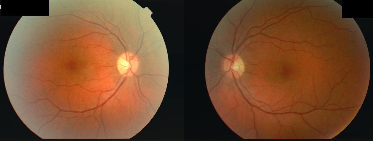
Figure 1. Retinal fundus photographs of the right and left eyes in a patient with acute optic neuritis, a common presenting manifestation of multiple sclerosis.
Acute optic neuritis (ON) is a common first symptom for a later diagnosis of multiple sclerosis (MS). It is known that high-contrast visual acuity—the ability to read black letters on a white background—tends to recover well after unilateral ON. However, low-contrast visual acuity (LCVA) recovery in patients with MS is less well known, whether affected or unaffected by ON, and Dr. Charmaine Yam and colleagues at Queen Square Institute of Neurology of University College London sought to examine LCVA. The team hypothesized that advanced MS would lead to worse color vision and LCVA in the long term, which would correlate with damage observed on optical coherence tomography (OCT) and magnetic resonance imaging (MRI).
Yam and colleagues examined 178 adults approximately 15 years after their first demyelinating event, using OCT and brain MRI to measure structural damage and brain volume, alongside high-contrast, low-contrast, and color vision assessments. Then, they used statistical models to determine whether long-term visual outcomes were associated with an MS subtype, optic neuritis, and markers of neurodegeneration.
The team found that eyes with a history of optic neuritis displayed worse color vision and reduced LCVA compared to unaffected eyes. Among patients with more advanced subtypes or stages of MS, even without a history of optic neuritis, their eyes demonstrated slight visual deficits. OCT scans showed thinner nerve layers, which were strongly correlated with poorer color and low-contrast vision, indicating the presence of lasting structural damage. MRI scans also revealed greater brain lesion burden, or lower total brain volume, and brain atrophy, which were likewise associated with poor visual acuity. However, the OCT scans were stronger predictors of early and localized neurodegeneration affecting vision than MRI scans.
Yam and colleagues’ findings highlight OCT as an alternative, more cost-effective marker of MS. Even in patients with no history of optic neuritis, changes in color vision are evidence of underlying neurodegeneration in the retina and brain, corresponding to the retinal thinning detected on OCT. The effectiveness of OCT can be improved by comparing different types of color vision tests to identify the most sensitive one in detecting nerve damage. Using a larger variation of patients with MS, or even expanding to other demyelinating diseases, can firmly establish OCT as a definitive test in tracking MS. Together, these findings suggest that OCT, combined with color vision testing, can make MS detection easier and more efficient, even before obvious symptoms appear, allowing patients to seek treatment earlier.
Work’s Cited:
[1] C. Yam, et al., Investigating colour vision and its structural correlates 15 years following a first demyelinating event. Journal of Neurology, Neurosurgery, and Psychiatry 96, 435–442 (2025). doi: 10.1136/jnnp-2024-334551
[2] Image retrieved from: https://commons.wikimedia.org/wiki/File:Optic_Neuritis.png


