
By Adil Ali Khan, Class of 2029
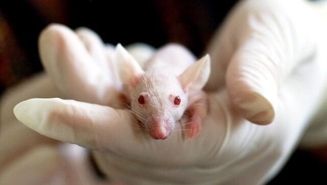
Figure 1: A male albino rat
Melanocytes in the hair matrix produce melanin, which gives hair its color. The various color combinations within their fur, which depend on the distribution pattern of hair follicles containing melanocytes, give rise to the coloration patterns observed in most terrestrial mammals. Albino rats do not genetically produce melanin, leaving their coats white, but researchers at Azabu University in Japan hypothesized that melanocytes differentiate and localize within these rats’ hair matrix, highlighting that pigment cells may be present but nonfunctional, an insight necessary to interpret melanocyte-related gene expression studies.
Tyrosinase helps convert tyrosine to melanin. Dopachrome tautomerase (Dct) is an enzyme commonly used as a specific marker for melanocyte lineage detection. Albino rats are often used as laboratory animals due to a mutation in the tyrosinase gene, from which they get their distinctive white coat. Otherwise, they are not considered to differ in any obvious way from wild-type pigmented rats. The researchers speculated that albino melanocytes do not produce pigment in the hair matrix. The researchers hypothesized that albino rats possess functional melanocytes capable of producing pigment, but that the tyrosinase mutation blocks the final step, leaving the coat white despite the cells being present.
For the experiment, researchers examined four rat strains: one pigmented (black) and three with albinism, and collected skin samples from different body regions. Fluorescence immunohistochemistry was used to locate melanocytes in hair follicles, and tyrosinase and Dct gene expression were measured to demonstrate the presence of melanocytes in the skin. The results showed that melanocytes are present even in the absence of observable pigment, as the tyrosinase and Dct genes were expressed in all three albino rats. Microscopy confirmed the presence of melanocytes in the hair follicles of albino rats, although no melanin was produced. Additional analysis revealed that the distribution of melanocytes varies across body parts, with some areas containing more melanocyte-containing follicles than others. However, a key limitation of this study is that only male albino rats were used, leaving the melanocyte distribution in female albino rats unexplored.
Overall, the results support the hypothesis that albino rats lack pigment not because melanocytes are absent, but because tyrosinase is defective, preventing melanin production. This research is significant because it provides the first documented report of melanocyte presence and distribution in the hair follicles of albino rats. These findings emphasize how a single gene can block pigmentation even when the cellular apparatus is intact and highlight that further research on female albino rats could provide additional insight.
Work’s Cited:
[1] Numata, N., Ozawa, A., & Sakaue, M. (2025). Regional differences in the distribution of melanocyte-containing hair bulbs in the skin of male albino rats. PloS one, 20(11), e0336110. https://doi.org/10.1371/journal.pone.0336110
[2] Image retrieved from: https://www.rawpixel.com/image/5949313/free-public-domain-cc0-photo

