

Mariam Malik ‘22
Cartilage is malleable soft tissue meant to prevent excess friction from joints rubbing against one another. Located in numerous parts of the body, such as the larynx, respiratory tract, and the septum, human cartilage is the most malleable and widespread at birth, eventually being replaced by bone. For a long time, scientists believed that cartilage in joints could not be regrown; however, in a recent study at Duke University Medical Center, researchers found that humans are indeed capable of regrowing cartilage, similar to the way that salamanders and zebrafish regenerate limbs.
A team of researchers under senior author Virginia Byers Kraus, professor in Duke’s Departments of Medicine, Pathology, and Orthopedic Surgery, along with lead author Ming-Feng Hsue, were aware from previous research that the ability of certain mammals to regenerate entire limbs is because of the presence of microRNA (miRNA) and the rate of protein turnover that inevitably occurs with tissue growth. In order to analyze the rate of protein turnover in groups of salamanders and zebrafish, the team used internal molecular clocks because cartilage proteins are long lived and thus prone to age-related amino acid alterations. Furthermore, it was crucial to understand that newly-made proteins have few to no amino acid conversions, whereas older proteins will have several alterations. The researchers used sensitive mass spectrometry and proteins altered by deamidation, which is the removal through hydrolysis of the protein’s amide group. Despite the abundance of glutamine throughout the body, the team used asparagine (Asn, N) as the deamidated protein because it is the only one that has dependable predictions of deamidation rates based off 2D and 3D protein features. Asn’s deamidation rates are also much faster than those of glutamine. By comparing the half-lives of protein, the team learned that the age of cartilage differs depending on where it is located in the body. Cartilage in the ankles, for example, has a shorter half-life than cartilage in the knee or hip.
Kraus and Hsue were aware that for animals with limb-regenerative capacity, blastema formation at the site of the limb injury is essential; therefore, they hypothesized that because miRNA is vital for blastema formation, it must also be involved in cartilage protein turnover. To determine whether human cartilage shares this quality, RNA was extracted from human ankle, knee, and hip cartilages to be analyzed for expression of miRNA, specifically the type that forms blastemas. The researchers found that miRNA concentration was the highest in ankles, compared to knees and hips, and higher in the first few layers than ones that were deeper.
The finding provides information into the potential of human cartilage to regenerate, as well as potential medicine made with miRNA that could stop, treat, or hinder arthritis. Kraus also adds the possibility of further research in figuring out which regulators salamanders have that humans do not.
References:
- M.F. Hsueh, et al., Analysis of “old” proteins unmasks dynamic gradient of cartilage turnover in human limbs. Science Advances 5, (2019). doi: 10.1126/sciadv.aax3203
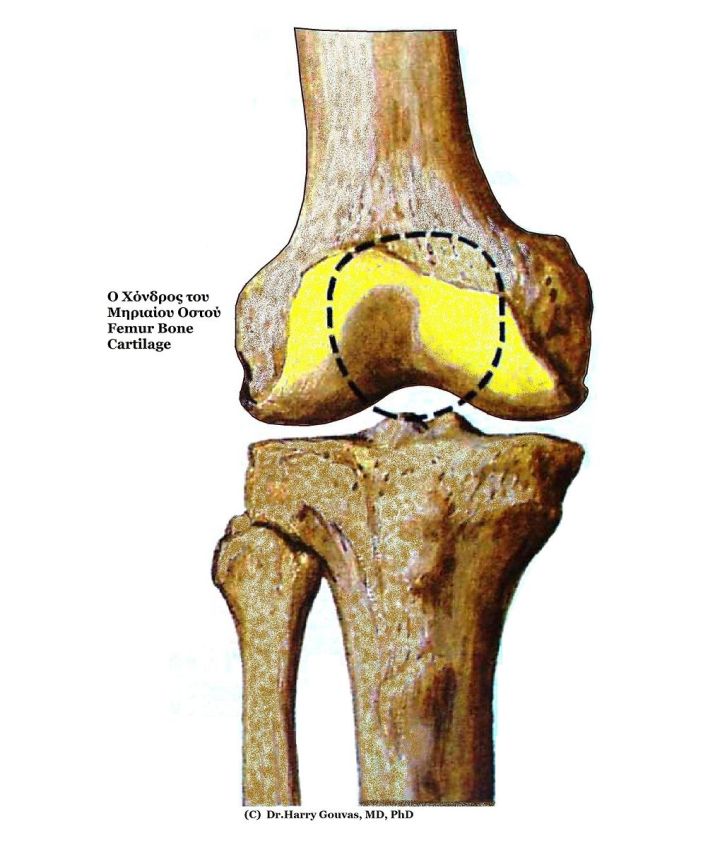
- Image retrieved from: https://commons.wikimedia.org/wiki/File:Knee_Femur_Cartilage.jpg
