
Author: Diego Javier, Class of 2026
Muscle regeneration is a nuanced and important area of study in understanding how muscles recover from injury. Researchers have been trying to develop therapies for muscle-related diseases, and some have used zebrafish as a model organism given their ability to regenerate their heart and skeletal muscles. In order to study muscle regeneration. Eric Paulissen, in the lab of SBU professor Benjamin Martin, sought to generate a new model by engineering zebrafish to express the enzyme nitroreductase (NTR) in skeletal muscle cells. Upon treatment with the drug metronidazole (MTZ), NTR converts MTZ into a chemical that causes muscle cells to undergo apoptosis (cell death), serving as a model for studying muscle regeneration. This targeted cell death allows researchers to study how zebrafish muscles regenerate after injury, making it a controlled model for investigating muscle repair mechanisms.
To create this transgenic zebrafish model, the researchers inserted a codon-optimized NTR gene under the control of a muscle-specific promoter. After confirming that MTZ treatment effectively killed muscle cells without damaging surrounding tissues, they tagged the enzyme with a fluorescent protein, which allowed them to visualize muscle cell death and regeneration under a microscope.
The researchers found that muscle tissue was largely ablated after MTZ treatment. After removing the drug, it was found that the zebrafish embryos began regenerating muscle fibers over several days. Time-course imaging had revealed that new striated muscle cells gradually appeared, and much of the skeletal muscle had recovered after 96 hours. In situ (in its place) hybridization experiments showed that muscle cells reentered a state of muscle formation, producing new differentiated fibers and showcasing the regenerative capacity of zebrafish.
Their findings also revealed how dying muscle cells interact with the immune system. Specifically, apoptotic cells attracted macrophages, which are immune cells that clear cellular debris and likely assisted with the regenerative process. Aside from natural regeneration, this zebrafish system allows for selective transplantation of transgenic muscle cells into wild-type embryos. This feature allows researchers to conduct controlled experiments of muscle repair in specific regions, with this being a powerful tool to investigate the dynamics of regeneration and the roles of different cell types in muscle recovery.
This transgenic zebrafish model provides a precise and reversible method for removing skeletal muscle, allowing for detailed study of various disciplines such as regeneration, immune interactions and tissue recovery. This model will help aid in future studies of muscle regeneration and can help contribute to future therapies for muscle damage and disease.
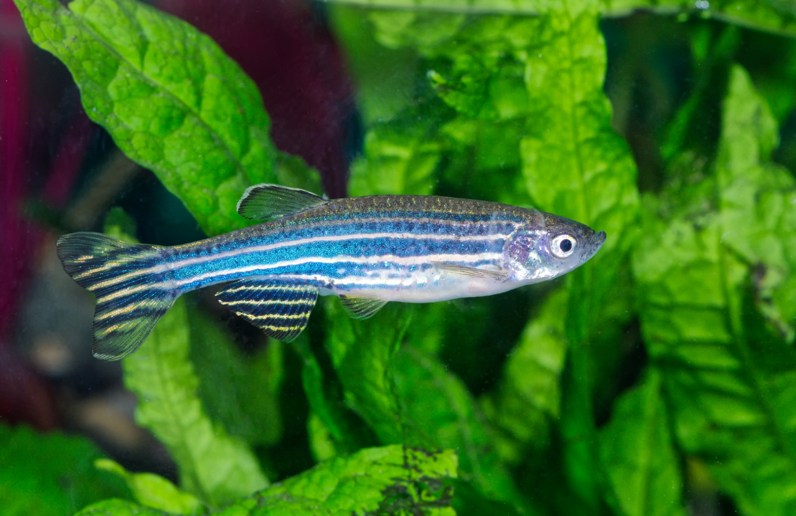
Figure 1. Zebrafish (Danio rerio), the model organism used in the study to investigate skeletal muscle ablation and regeneration.
Works Cited:
[1] Paulissen, E., & Martin, B. L. (2024). A Chemically Inducible Muscle Ablation System for the Zebrafish. Zebrafish, 21(3), 243–249. https://doi.org/10.1089/zeb.2023.0102
[2] Image retrieved from: https://www.understandinganimalresearch.org.uk/using-animals-in-scientific-research/animal-research-species/zebrafish
