

Julia Chivu ’23
Three dimensional culture systems and patient derived cells successfully allow for tumoroid development. Tumoroids are structures that grow and appear morphologically similar to naturally growing tumors in a patient. Tumoroids show promise for testing new drugs and cancer treatments. For instance, glioblastoma is a form of brain cancer that is aggressive, fast growing, and deadly. In particular, the mesenchymal subtype of this cancer shows more proliferative growth in a faster period of time. To better understand the tumoroid development, Dr. Lisa Oliver and her research team have created a 3D cell culture model, which mimics the environment in which glioblastoma and mesenchymal tumor cells grow.
To mimic the tumor microenvironment found in the brain, a co-cell culture was made using patient derived cells and bone marrow mesenchymal stem cells. A co-culture refers to a cell culture that contains two or more types of cells. The patient derived cells were collected from patients that were undergoing glioblastoma tumor removal surgery. The stem cells were collected from a French tumor library. The cell viability, cell proliferation, and percentage of tumor-initiating cells was analysed for the growing tumoroids. The morphology of the tumoroids, such as their length, area, and circularity, were analyzed. Additionally, radiation and chemotherapy were used on these tumoroids to determine their survival and cell proliferation probabilities.
The researchers noticed that patient derived cells began to create multicellular spheroids within seven days of being placed in the co-culture. It was inferred that the tumoroids responded to radiation methods, as there was a decrease in the number of cells in the culture. In addition, the morphology and behavior of the lab-grown tumors exhibited similar characteristics to patient-related glioblastoma and mesenchymal tumors.
It is believed that this new procedure could allow for the development of an enhanced tumor microenvironment in comparison to that of a two dimensional petri dish cell culture. The research group hopes that their model can be used to individualize treatment options for those diagnosed with glioblastoma. This research is vital in learning more about the components and functions of tumor formation. In addition, these findings may play a significant role in the development of new cancer therapeutic treatments.
Works Cited:
[1] L. Oliver, et al., A Simple 3D Cell Culture Method for Studying the Interactions Between Human Mesenchymal Stromal/Stem Cells and Patients Derived Glioblastoma. Cancers 15, 1304 (2023). doi: 10.3390/cancers15041304
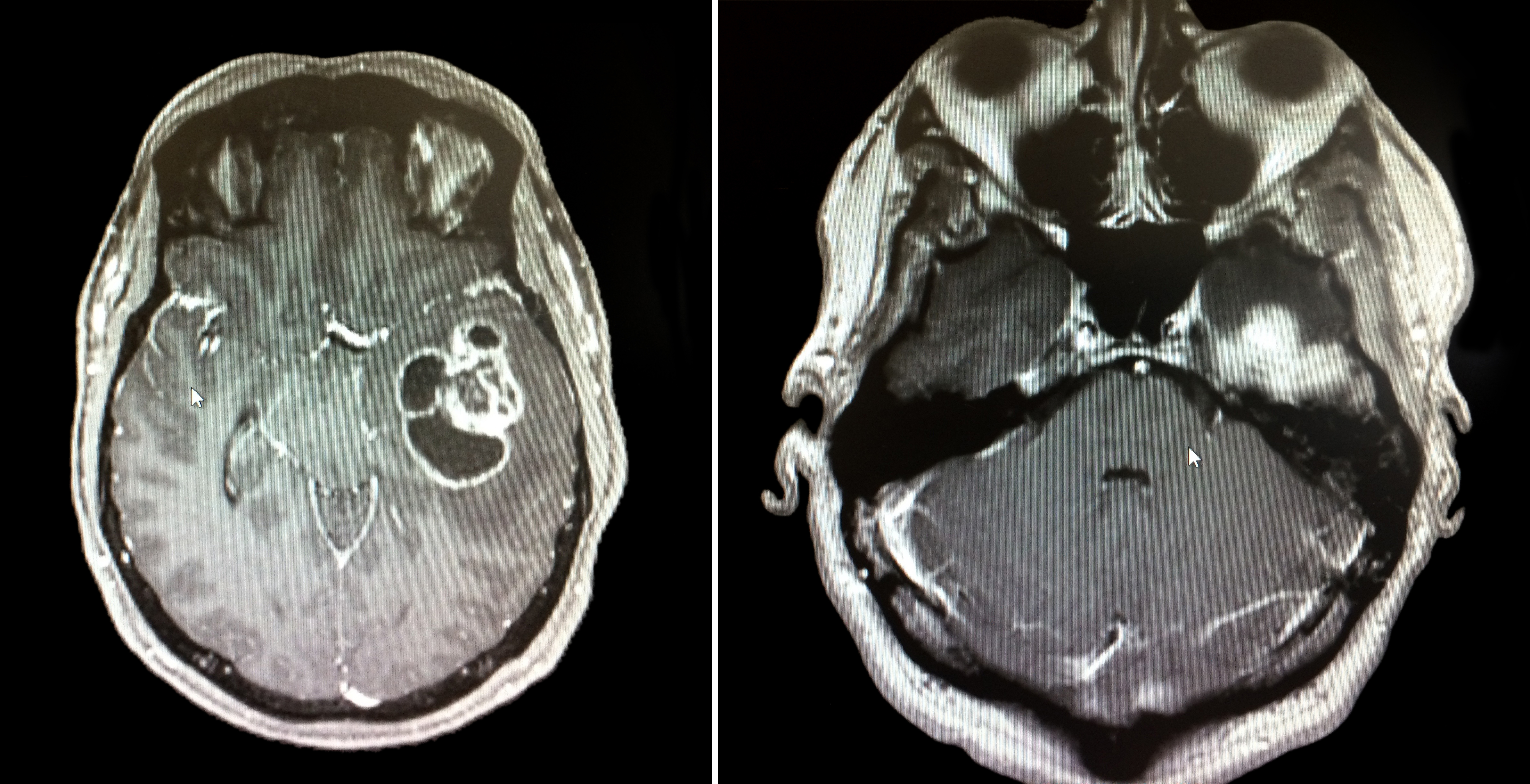
[2] Image retrieved from: https://www.flickr.com/photos/cambridgeuniversity-engineering/48664106968/in/photolist-2h9hgz3-2h7PqWH-oy9f2r-oeHot9-oeWFYq-wM5iPn-oepNiP-brPSi4-gEmw7w-pWR3uF-oeMRTx-owKCS8-oujHTR-wZraKH-oeWG8o-ovFJVq-24KtuT-ousAEM-x395yf-oubVmw-oezokj-xH7qHd-oxCbBF-oeWJN3-x1zisY-gu2Fug-wL9Sz7-oeWDRN-2h7MxsV-x4pYNt-oxPgkM-ow8vYt-ouiMAc-ovzjrK-ouc4Kq-2gh4edq-oetLao-oeJc2P-w6NQC3-cmWB6-oeByDa-275VWGW-owvWXp-otPDMC-w6W4YM-ow3h1m-ovMQXR-wLaut1-owmAp7-owR2ya
