

Vignesh Subramanian ’24
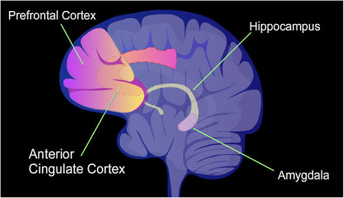
Childhood adversity is a broad term encompassing a range of experiences and circumstances that negatively impact a child’s well-being and development. Such circumstances include trauma and exposure to violence, all forms of abuse, neglect, exploitation, and family economic hardship, with profound and stratified implications for lifelong health. Lasting effects often include the development of toxic stress responses, accumulating hormones that over-activate certain immune and metabolic reactions and placing the body in a prolonged “fight or flight” state without full recovery. These responses exhaust the body and drive internal anatomical damage, particularly to the architecture of the developing brain and vasculature. The physiological consequences for brain structures involved in regulating said stress— namely, the amygdala, hippocampus, and prefrontal cortex (PFC)— have been increasingly studied in recent years, with particular focus on differences in their development among children of different racial and socioeconomic backgrounds.
Researchers led by Dr. Harnett of Harvard University worked to characterize the relationship between racial disparities in childhood adversity and neuroanatomical differences. The researchers first sampled 7,350 White and 1,786 Black American children from the ongoing Adolescent Brain Cognitive Development (ABCD) study, collecting their family demographic data, ranking their neighborhoods by socioeconomic disadvantage with a standardized index, and assessing the family conflict, material hardship, and trauma history they faced using questionnaires. The researchers then collected structural MRI data at 21 points across 14 regions of interest in the gray matter of the children’s brains, applying statistical analyses to correlate gray matter volume with the indices of adversity collected earlier.
The researchers found that, overall, greater exposure to the family- and trauma-linked adversities investigated was linked to lower gray matter volumes in the amygdala, hippocampus, insula, and nearly a dozen subregions of the PFC. Accordingly, Black children showed lower gray matter volumes in all of these regions compared with White children. Statistically accounting for these racial disparities in adversity exposure resolved race-related neuroanatomical differences in some of these regions, strongly suggesting said disparities to be primary contributing factors. These findings align with the preexisting understanding that the afflicted regions undergo rapid development early in childhood and at times are strongly impacted by concentrated stress responses (though those impacts may be either immediate or delayed). Future research may build upon this study by incorporating other racial groups and longitudinal MRI scans rather than merely cross-sectional data.
Works Cited:
[1] N. Harnett, et al., Racial Disparities in Adversity During Childhood and the False Appearance of Race-Related Differences in Brain Structure. American Journal of Psychiatry 180, 127-138 (2023). doi: 10.1176/appi.ajp.21090961
[2] Image retrieved from: https://commons.wikimedia.org/wiki/File:PTSD_brain.svg
