

Vignesh Subramanian ’24
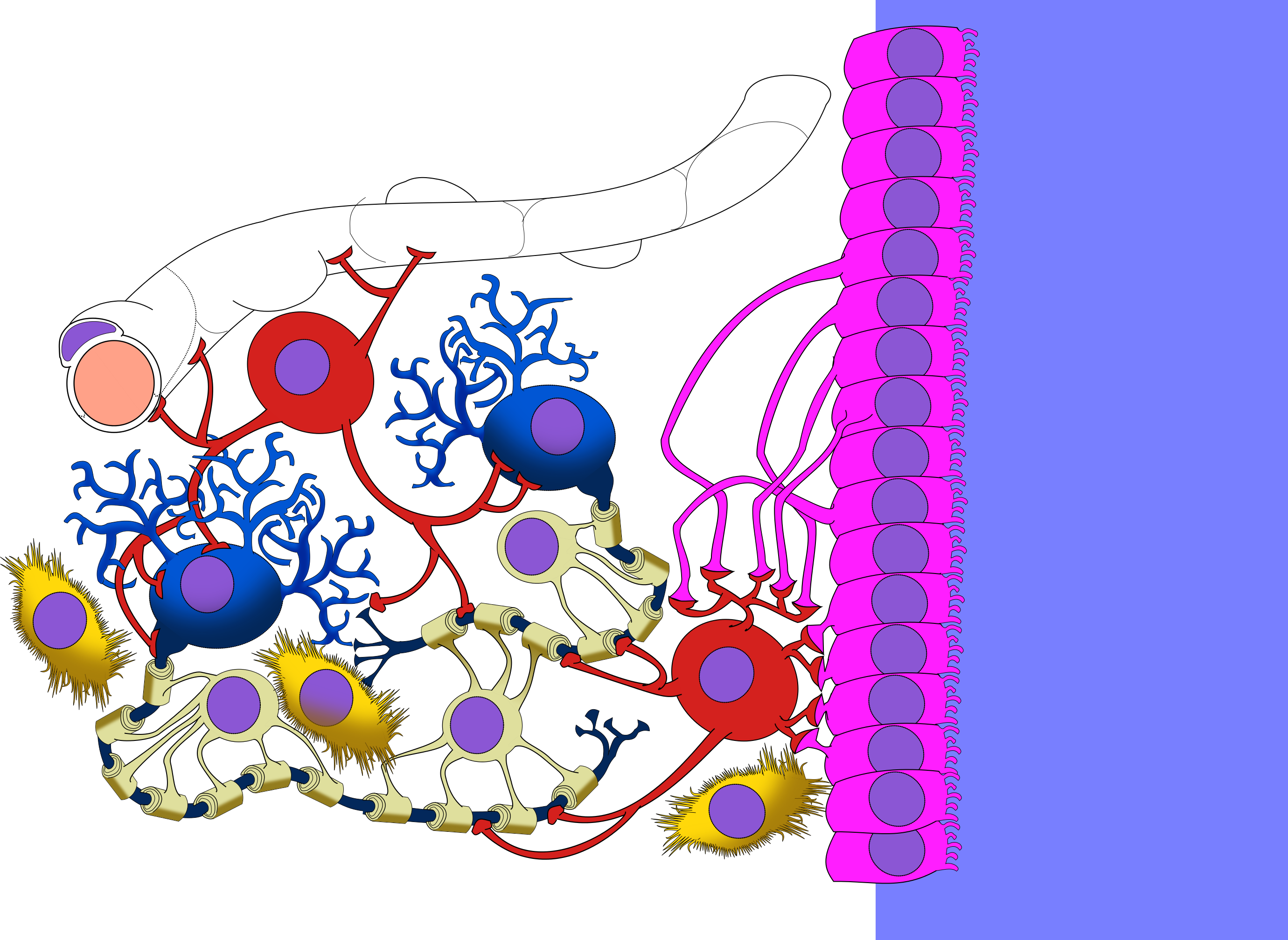
Glia are non-neuronal cells that host and provide a number of homeostatic ancillary functions in both the central (CNS) and peripheral (PNS) nervous systems. Though only recently discovered and characterized, glial cells vastly outnumber neurons and provide them with structural support and insular protection, driving their myelination (the process of forming coverings known as myelin sheaths that coat neurons’ signaling axons), facilitating their communication, and transporting their waste. Specialized glia such as oligodendrocytes, which myelinate CNS neurons, are differentiated from a rare glial cell type known as neuron-glial antigen 2 (NG2)-expressing glia. Though they primarily serve as precursors, NG2 glia are also thought to be involved in promoting the uptake of excitatory neurotransmitters such as glutamate that may counteract the effects of chronic stress. A study led by Dr. Adan Aguirre of Stony Brook University aimed to better understand the relationship between NG2 presence in the brain and stress-associated behavioral phenotypes.
The researchers first administered intraperitoneal injections of diphtheria toxin (DT) to a transgenic adult mouse line engineered to place DT receptors under the control of the NG2 gene promoter, enabling DT administration to induce death of NG2 glia. Following visualization of NG2 glia ablation, or selective destruction, in the prefrontal cortices (PFCs) of the mouse brains, miniature excitatory postsynaptic currents from pyramidal neurons in PFC slices were recorded to assess postsynaptic excitatory glutamate receptor function. Genetic knockdowns of extraneous signaling models, mRNA expression quantification, and congenital models of maladaptive responses were then used to isolate and identify stress-induced depressive and anxious behavior phenotypes in the mice.
The researchers found that NG2 glia direct the production of a growth factor (FGF2) that stimulated other glia (e.g., astrocytes) to regulate glutamate receptor trafficking and synaptic presence, as well as glutamate uptake, establishing a relationship between capacity for excitatory neurotransmission and NG2 glia presence. Decreased FGF2 secretion was found to cause brain region-specific deficits in glutamatergic signaling, such as those of the PFC, that were correlated with depressive-like behaviors such as learned helplessness being exhibited following genetically-induced NG2 glia ablation. Together, these findings directly link cell NG2 glial cell density and depletion to stress symptom emergence, revealing a pathway by which similar human brain pathophysiology implicated in untreated mood disorders, such as major depressive disorder (MDD), may be isolated and therapeutically targeted.
Works Cited:
[1] A. Aguirre, et al., Genetic and Stress-Induced Loss of NG2 Glia Triggers Emergence of Depressive-Like Behaviors through Reduced Secretion of FGF2. Neuron 88, 941-956 (2015). doi: 10.1016/j.neuron.2015.10.046
[2] Image retrieved from:
https://commons.wikimedia.org/wiki/File:CNS_glia.png
